By which of the following mechanisms can a cell transport a substance from a lower to a higher concentration. Streptococcus contains a variety of species.

Chapter Four Microbiology Flashcards Quizlet
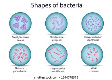
Streptococci are spherical organisms that grow in chains because of incomplete separation after division of the cells Figure 1They were first described in 1874 by Billroth who used the term Streptococcus from two Greek words.
. Learn vocabulary terms and more with flashcards games and other study tools. 72Antibiotics that target cell wall synthesis ultimately cause bacterial. Streptococci are microbiologically characterized as gram-positive and nonmotile.
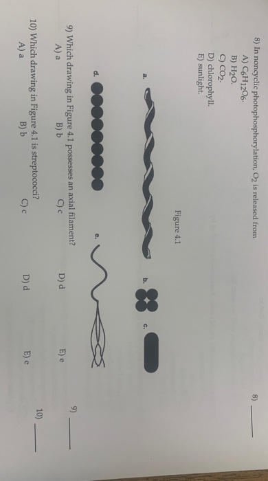
Start studying Chapter Four Microbiology. Streptococci illustration figure drawing diagram image. Which drawing in Figure 41 possesses an axial filament.
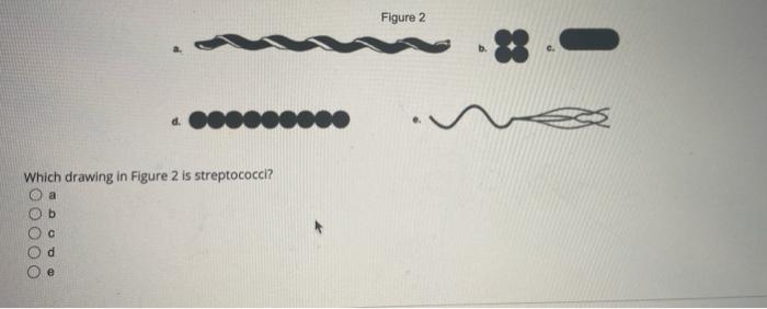
Figure 2 which drawing in figure 2 possesses an axial. Streptococcus Bacteria Classification Shape Infection Gram Stain Overview. When incubated aerobically this group of streptococci may render less obvious β-hemolysis also known as α-prime hemolysis which is.
C CO2 D chlorophyll. 28 28 In Figure 41 which drawing represents a spirochete bacterium with an axial filament. А а B - Cc D E e 10 Which drawing in Figure 41 is streptococci.
____ Which of the following statements about prokaryotic cells is generally false. In group A streptococci the R and T proteins may serve as epidemiologic markers but the M proteins are clearly virulence factors associated with resistance to phagocytosis. Streptococcus pyogenes are spherical to ovoid microorganisms measuring up to 1 μm in diameter.
Which of the following statements about gram-negative cell walls is FALSE. D neither a nor b. Streptococcus bacteria is Gram-positive and are generally spherical in shape.
27 In Figure 43 which diagram of a cell wall is a gram-negative cell wall. Some strains may produce mucoid colonies Figure 1. Which drawing in the figure is streptococci To apply just stick on your nails we like to keep them at The bottom of our nail beds and set with clear topcoat.
More than 50 types of S pyogenes M proteins have been identified on the basis of antigenic specificity. They are commonly found in the mucous membrane of the mouth and respiratory tract etc where they have been associated with a number of diseases and infections including sepsis pneumonia and pharyngitis. A a B Б C с streptococci Cc da te.
Which drawing in Figure 41 is streptococci. This preview shows page 4 - 7 out of 9 pages. Both the M proteins and lipoteichoic.
20 In Figure 43 which diagram of a cell wall is resistant to many antibiotics eg penicillin. The cell wall also consists of several structural proteins Figure 13-2. Agalactiae colonies can be flat grayish-white or orange mucoid and creamy.
Which drawing in Figure 41 is a tetrad. A a B b C both a and b D neither a nor b E The answer cannot be determined based on the information provided. E The answer cannot be determined based on the information provided.
A a B Б C с streptococci Cc da te. The resolution of a microscope can be improved by changing the A. The term streptococcus twisted berry refers to the bacterias characteristic grouping in chains that resemble a string of beads.
PowerPoint Win Mac compatible. Which drawing in Figure 41 is streptococci. C 9 Which drawing in Figure 41 possesses an axial filament.
Which drawing in Figure 41 is a bacillus. 8 In noncyclic photophosphorylation O2 is released from A C6H1206 B H20. Streptococci are facultatively anaerobic Gram-positive organisms that often occur as chains or pairs figures 1 and 2 and are catalase-negative in contrast staphylococci are catalase positive figure 3.
____ Which of the following statements about prokaryotic cells is generally false. Question 32 1 1 pts Which drawing in Figure 41 is streptococci Figure 41 D d from SCIENCE 290 at West Coast University Los Angeles. A a B b C c D d E e 29 Bacillus and 29 In Figure 41which drawing represents the shape of endospore-producing Clostridinen A a B b C c D d E e 30 30 Which enzyme catabolizes fats into glycerol and fatty acids.
C both a and b. Streptococci are subdivided into groups by antibodies that recognize surface antigens figure 4. 71Which drawing in Figure 41 is streptococci.
A axial filament b tetrad c single bacillus d streptococci e. 69Which drawing in Figure 41 is a tetrad. Which drawing in Figure 41 possesses an axial filament.
They possess 80S ribosomes. 70Which drawing in Figure 41 possesses an axial filament. Figure 2 Which drawing in Figure 2 possesses an axial filament.
A watch-catching style has never been less complicated. This illustration is included in the following Illustration Toolkit. Wavelength of light to shorter wavelenght D.
Which drawing in Figure 41 is streptococci. They possess 80S ribosomes. Which drawing in Figure 41 is a tetrad.
Extracellular enzymes Facilitated diffusion integral. In the beginning streptococci were classified according to the disease they caused. Which Drawing In The Figure Is Streptococci.
Which drawing in Figure 41 is a bacillus. True False Which drawing in the figure is streptococci b e a d c Which drawing from BIOL 2304 at Lamar University. Streptos chain kokhos berry.
Streptococcus genus Streptococcus group of spheroidal bacteria belonging to the family Streptococcaceae. Antibiotics that target cell wall synthesis ultimately cause bacterial cell death as a result of.
Solved Figure 2 D Which Drawing In Figure 2 Is Chegg Com
Solved 8 In Noncyclic Photophosphorylation O2 Is Released Chegg Com

2 1 Sizes Shapes And Arrangements Of Bacteria Biology Libretexts Bacteria Prokaryotic Cell Bacterial Cell Structure

Chapter 4 Questions Flashcards Quizlet
Streptococci Images Stock Photos Vectors Shutterstock

Chapter 4 Practice Quiz Flashcards Quizlet

Chapter 4 Questions Flashcards Quizlet

Functional Anatomy Of Prokaryotic And Eukaryotic Cells Ppt Video Online Download
0 comments
Post a Comment